Keratoconus is a condition in which the cornea, the clear front surface of the eye, becomes thinner and bulges forward, causing blurred and distorted vision. It often starts in the teens or 20s, which is why many young patients in India are diagnosed during college or early working years. The two big goals of treatment are to improve vision and to stop the cornea from worsening. Modern options include glasses, specialty contact lenses, corneal cross-linking, ring procedures, selected laser or lens-based treatments, and in advanced cases, corneal transplant surgery.
Keratoconus – Causes, Stages & Modern Treatment (C3R/CXL)
Keratoconus is one of the most important corneal conditions I explain to young patients and their families. It often begins at an age when people are studying, using screens for long hours, preparing for exams, learning to drive, or starting work. Indian data show that keratoconus commonly presents in the second and third decade of life, with a median age at presentation around 22 years. That is why early diagnosis matters so much.
The good news is that keratoconus is no longer a condition we simply “watch and wait” until vision becomes very poor. Today, we have better corneal scans, better specialty contact lenses, and a treatment called corneal collagen cross-linking, also known as CXL or C3R, that can slow or stop progression in many patients. We also have more options for advanced disease than before.
What is keratoconus?
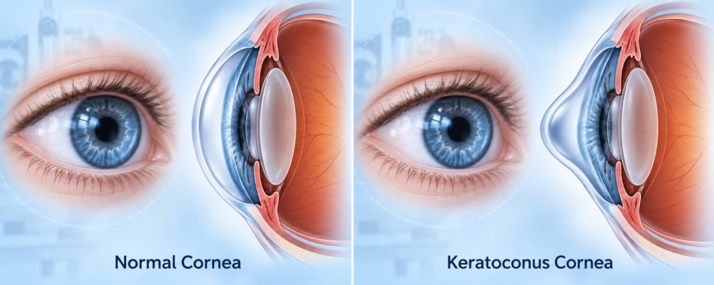
The cornea is the clear, dome-shaped front window of the eye. In a healthy eye, it has a smooth and regular curve that helps focus light sharply on the retina. In keratoconus, the cornea gradually becomes thinner and bulges outward into a cone-like shape. Because the front surface is no longer smooth, light does not focus properly, and vision becomes blurred, stretched, or shadowed. Keratoconus usually affects both eyes, but one eye is often worse than the other.
Patients often describe this in very practical ways. They say the power in their glasses keeps changing, headlights scatter at night, letters seem doubled, or one eye feels much weaker than the other. That is typical of keratoconus because the issue is not only the number in the glasses. The shape of the cornea itself is changing.
Why keratoconus happens
There is no single cause of keratoconus. It is usually understood as a condition caused by a mix of genetic tendency and environmental stress. Family history is important, and Mayo Clinic notes that about 1 in 10 people with keratoconus has a parent with the condition.
Some risk factors are seen again and again in clinic. These include frequent eye rubbing, allergy-related itching, eczema, asthma, and certain connective tissue or developmental conditions. Eye rubbing deserves special attention because it is one of the commonest habits linked with worsening disease, especially in young people with allergic eyes.
In simple terms, I tell patients this: if your cornea is already vulnerable, repeated rubbing adds extra mechanical stress. That does not mean rubbing is the only cause, but it is one of the few risk factors we can actually try to control.
Keratoconus symptoms patients should not ignore
Keratoconus symptoms usually develop gradually. Common symptoms include blurred vision, distorted vision, glare, halos around lights, increasing sensitivity to light, and repeated changes in spectacle number. Some patients notice ghost images or multiple outlines around text, especially in dim light.
One of the biggest clues is “changing astigmatism.” A teenager or young adult who keeps getting new glasses every year, especially with increasing cylindrical power, should not be dismissed as routine. In early disease, the cornea may still look fairly normal on basic examination, but the shape may already be becoming irregular.
In advanced cases, the cornea can become scarred. Some patients can develop a sudden painful episode called acute hydrops, where fluid enters the cornea and causes sudden clouding and drop in vision. It is not the most common presentation, but it is important because sudden worsening in a keratoconus patient needs prompt review.
How keratoconus is diagnosed
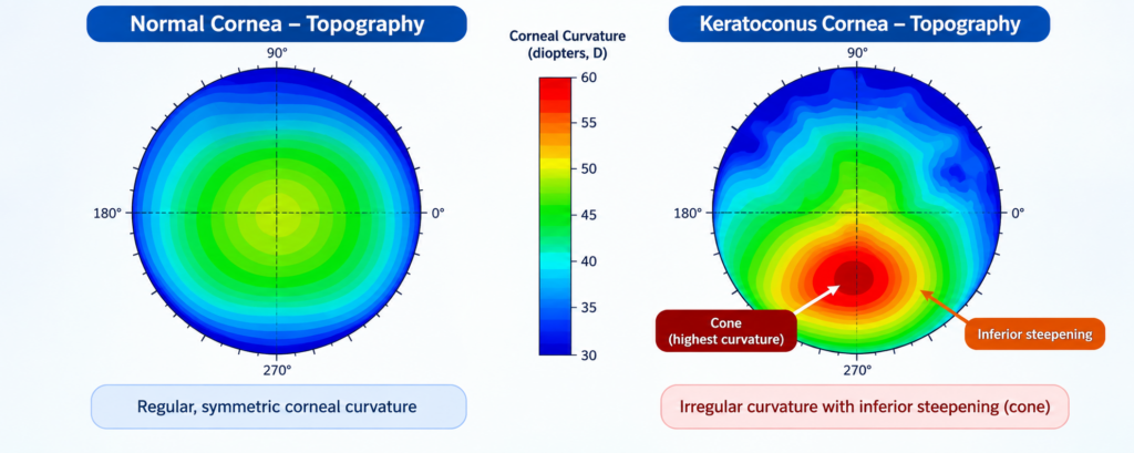
Keratoconus is diagnosed with a combination of refraction, slit-lamp examination, keratometry, and most importantly corneal imaging. The two most useful scans are corneal topography and corneal tomography. Topography maps the front surface shape of the cornea. Tomography goes further by evaluating the front surface, back surface, and thickness pattern, which is why it is especially useful for early or suspicious cases.
Modern diagnosis is much better than it was years ago because the back surface and thickness profile of the cornea can now reveal early disease before it becomes obvious on routine examination. That is one reason tomography has become so important in patients with irregular astigmatism, family history, or repeated power changes.
Understanding keratoconus stages
Patients are often told they have mild, moderate, or advanced keratoconus. These labels are useful, but modern staging has moved beyond simple front-surface steepness. Reviews of current staging explain that newer systems, such as the Belin ABCD classification, incorporate anterior curvature, posterior curvature, thinnest pachymetry, and visual acuity, giving a more complete picture of the disease.
For patients, I prefer a simpler explanation. In mild keratoconus, glasses or soft toric lenses may still work. In moderate disease, the cornea becomes more irregular and specialty contact lenses are often needed. In advanced disease, the cornea may become very steep, very thin, scarred, or difficult to manage with lenses, and surgery becomes more relevant. More important than a stage label is whether the scans are stable or getting worse over time.
The two goals of keratoconus treatment
Keratoconus treatment has two separate aims. One is to improve vision. The other is to stop progression. This is where many patients get confused. Glasses and contact lenses improve sight, but they do not strengthen the cornea. Cross-linking strengthens the cornea, but it does not necessarily remove the need for glasses or contact lenses. Different treatments solve different parts of the problem.
Keratoconus glasses and specialty contact lenses
In the earliest stages, keratoconus glasses may still provide reasonable vision. Soft toric contact lenses may also help while the cornea is still fairly regular. But once the cornea becomes more uneven, glasses usually stop giving crisp vision because they cannot fully neutralize irregular astigmatism.
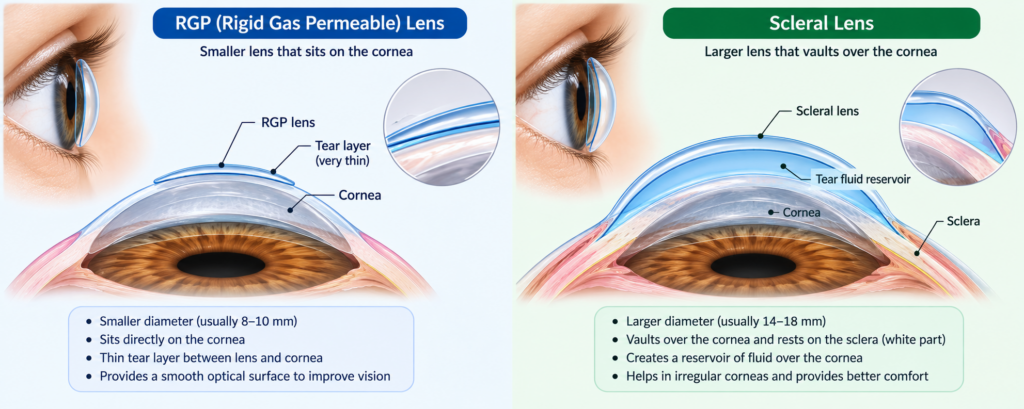
At that point, specialty lenses become very important. Rigid gas permeable, or RGP, lenses create a smooth optical surface over the irregular cornea. Hybrid lenses combine a rigid center with a softer edge. Scleral lenses are larger lenses that rest on the white part of the eye and vault over the cornea, with a fluid layer in between. Mayo Clinic notes that scleral lenses are a good alternative to surgery for many keratoconus patients.
This is a useful place to internally link to your hospital’s articles on RGP lenses, scleral lenses, and contact lens options for irregular corneas.
Corneal Collagen Cross-Linking (CXL)
Corneal collagen cross-linking, or CXL, is the most important modern treatment for progressive keratoconus. It uses riboflavin eye drops and ultraviolet-A light to stiffen the corneal collagen and help stop further bulging. The American Academy of Ophthalmology states that corneal cross-linking is the recommended treatment for progressive keratoconus because it stabilizes the cornea and reduces the risk of progressive ectasia.
Moorfields explains that CXL, also called C3R, is designed to prevent keratoconus from getting worse. That is the key message for patients. It is mainly a disease-stabilizing treatment, not a spectacle-removing treatment. After CXL, many patients still need glasses or contact lenses, but the aim is to protect the cornea from further deterioration.
Younger patients often benefit the most from timely CXL because they are more likely to show progression. The decision is usually based on age, change in topography or tomography, refraction change, and corneal thickness.
Intracorneal Ring Segments (ICRS/INTACS)
Intracorneal ring segments, also called ICRS or INTACS, are small synthetic arc-shaped implants placed within the cornea. Their role is to flatten and regularize the corneal surface in selected mild to moderate cases. Mayo Clinic includes them among treatment options that can help flatten the cornea, improve vision, and sometimes make contact lens fitting easier.
Patients should understand that ICRS do not replace the cornea and do not act like a transplant. They are a reshaping procedure. Reviews describe them as a useful option in carefully chosen patients who have meaningful irregularity but still have a reasonably clear central cornea and enough thickness for implantation. They may also be combined with CXL in some cases.
Corneal Allogenic Intrastromal Ring Segments (CAIRS)
CAIRS is a newer variation of ring-segment treatment. Instead of synthetic segments, it uses donor corneal tissue segments placed inside the cornea. This is a more specialized option and is usually offered in advanced corneal centers rather than as a routine first-line treatment.
Recent reviews and studies suggest that CAIRS can improve vision and corneal shape in selected keratoconus patients and may be a safe and effective option in experienced hands. For patients, the simplest way to understand CAIRS is that it is a tissue-based ring procedure used in selected cases when the surgeon feels it may offer better corneal reshaping or a useful alternative to synthetic segments.
Topography-Guided PRK with CXL (Athens Protocol)
Topography-guided PRK combined with CXL, often called the Athens Protocol, is one of the more advanced and selective options in keratoconus care. The idea is to use a customized surface laser treatment to make the front of the cornea smoother and then use cross-linking to strengthen it.
This is not a routine treatment for every patient with keratoconus because PRK removes tissue, and tissue removal must be used very cautiously in a cornea that is already weak. That is why the Athens Protocol is generally reserved for carefully selected eyes with enough thickness and a pattern of irregularity that may benefit from corneal regularization. Published reviews describe it as a promising strategy with long-term safety and efficacy in selected cases.
Phakic Intraocular Lens (ICL) Implantation
A phakic intraocular lens, or ICL, is a lens implanted inside the eye while the natural lens is left in place. In keratoconus, this is usually considered only after the cornea is stable, either naturally stable or stabilized with CXL, and when a patient still has a moderate to high glasses number that is not adequately managed with other methods.
This point is important: ICL does not treat the corneal weakness itself. It treats the remaining refractive error inside the eye. Reviews conclude that phakic IOL implantation can be a valid approach in stable keratoconus patients with moderate to high refractive error, especially when contact lens tolerance is poor and central scarring is absent.
Cataract Surgery with Toric or Customized IOL
Keratoconus patients can also develop cataract as they get older. Cataract surgery in these eyes needs extra planning because corneal measurements may be less predictable, especially when the astigmatism is irregular. A recent review notes that preoperative planning, IOL power calculation, and case selection are especially important in cataract surgery for keratoconus patients.
Toric or customized IOLs can be helpful in selected patients, particularly when the keratoconus is stable and the astigmatism is reasonably regular. Studies show that toric IOL implantation can improve vision and reduce astigmatism in suitable keratoconic eyes, but patients with very irregular corneas may still need glasses or specialty contact lenses even after a successful cataract operation.
Bowman Layer Transplantation
Bowman layer transplantation is a specialized surgical option usually considered in advanced keratoconus rather than early disease. It is not as commonly offered as CXL or standard transplant surgery, but it has an important role in selected advanced cases.
The goal is to strengthen and flatten the cornea enough to delay or avoid a larger transplant procedure. Reviews describe Bowman layer transplantation as a promising and relatively less invasive option for advanced progressive keratoconus in selected patients. In practical terms, I explain it as a “bridge” treatment that may help postpone full graft surgery in expert centers.
Deep Anterior Lamellar Keratoplasty (DALK)
DALK is a partial-thickness corneal transplant. In this procedure, the diseased front layers of the cornea are replaced, but the patient’s own healthy endothelium, the inner cell layer, is preserved. Mayo Clinic describes DALK as a transplant option used when the inner corneal layer can be kept.
For keratoconus, DALK is often preferred over full-thickness transplant when the endothelium is healthy because preserving that inner layer reduces the risk of endothelial rejection. Meta-analyses report fewer rejection-related problems and fewer postoperative complications with DALK compared with PKP, although visual recovery and surgical complexity can differ.
Penetrating Keratoplasty (PKP) – Full-Thickness Corneal Transplant
PKP, or penetrating keratoplasty, is a full-thickness corneal transplant. In this surgery, the entire cornea is replaced with donor tissue. Mayo Clinic describes PKP as a full-thickness cornea transplant used when complete replacement is required.
PKP is usually reserved for advanced keratoconus with severe scarring, extreme thinning, previous hydrops-related damage, or situations where lamellar surgery is not suitable. It can provide excellent vision in appropriate cases, but because donor endothelium is also transplanted, rejection risk is higher than with DALK. That is why many corneal surgeons prefer DALK when feasible and keep PKP for eyes that truly need a full-thickness graft.
Image suggestion: Simple illustration comparing DALK and PKP, showing partial-thickness versus full-thickness transplant
How doctors choose the right treatment
The easiest way to understand the treatment ladder is this. If the main problem is blur, we think about glasses or specialty contact lenses. If the main problem is progression, we think about CXL. If the cornea is irregular enough that reshaping may help, we may discuss ICRS, CAIRS, or in selected cases topography-guided PRK with CXL. If the cornea is stable but a large refractive number remains, ICL may be considered. If cataract is present later in life, cataract surgery with a toric or customized IOL may help selected patients. If the cornea is too scarred or too advanced, DALK or PKP enters the picture.
This is why no doctor should promise one “best” keratoconus treatment to every patient. The right treatment depends on age, scan progression, thickness, scarring, contact lens tolerance, and day-to-day visual needs.
A practical note on keratoconus surgery cost
Many people search online for keratoconus surgery cost, but keratoconus management is not one single surgery. One patient may only need glasses and monitoring. Another may need CXL. Another may need specialty lenses. Another may need ring procedures or transplant surgery. Because the treatment range is so broad, cost varies widely depending on the stage of disease, the technology used, whether a specialty lens fit is needed, and whether surgery is performed in a day-care setting or an operating theatre. This is an inference based on the wide range of accepted treatment pathways described in major clinical sources.
What patients can do to protect their vision
The first and most practical advice is simple: stop rubbing your eyes. If allergies are causing itching, get the allergy treated instead of rubbing through it every day. That small change can matter.
The second rule is not to skip follow-up. Keratoconus is often monitored by repeat scans, not just by symptoms. A patient may feel roughly the same, yet tomography can show progression. That is especially important in younger patients.
The third rule is to think about family screening when appropriate. Because family history is a known risk factor, siblings or children with frequent power changes, eye rubbing, or allergy symptoms should not ignore them.
When should you see an eye doctor?
Please get a corneal evaluation if your glasses number changes frequently, your astigmatism keeps increasing, night glare is becoming difficult, or one eye is much blurrier than the other. These are common real-life clues that simple refraction may not be telling the whole story.
Seek urgent care if there is a sudden drop in vision, sudden corneal clouding, or significant pain in a keratoconus eye. Sudden change is not something to wait on.
Final takeaway
If I had to summarize keratoconus in one sentence, I would say this: it is a corneal shape problem that needs timely diagnosis, careful monitoring, and stage-appropriate treatment. The earlier it is detected, the more likely we are to preserve useful vision with glasses, specialty lenses, and cross-linking before the disease becomes advanced.
Modern keratoconus care is far more complete than many patients realize. We now have a treatment ladder that includes corneal collagen cross-linking, intracorneal ring procedures, CAIRS, the Athens Protocol in selected cases, phakic ICL implantation, cataract surgery with toric or customized IOLs, Bowman layer transplantation, DALK, and PKP. The key is matching the right treatment to the right cornea at the right time.
Book an appointment with our cornea specialist for a detailed keratoconus evaluation, corneal topography, and a treatment plan tailored to your stage, scans, and visual needs.
References
American Academy of Ophthalmology. Corneal Ectasia Preferred Practice Pattern, 2024
Mayo Clinic. Keratoconus: Symptoms and causes
Mayo Clinic. Keratoconus: Diagnosis and treatment
Mayo Clinic. Cornea transplant
Moorfields Eye Hospital. Corneal cross-linking
Keratoconus: An updated review
Keratoconus in India: Clinical presentation and demographic distribution based on big data analytics
ABCD: A new classification for keratoconus
Corneal Allogenic Intrastromal Ring Segments: A Literature Review
Phakic intraocular lens implantation in keratoconus patients
Cataract surgery in eyes with keratoconus: A review of the current literature
Bowman Layer Transplantation for Treating Keratoconus: Preliminary Findings