Superficial / Automated Lamellar Keratoplasty in Hyderabad: Cost, Procedure & Recovery
Superficial / Automated Lamellar Keratoplasty is a specialized eye surgery designed to restore clear vision by replacing only the damaged front layers of your cornea. This advanced procedure carefully removes and replaces the diseased outer corneal tissue, preserving the healthy deeper structures of your eye. It’s a precise way to treat various corneal conditions, helping you see better and more comfortably.
QUICK FACTS
- Procedure Duration: 30-60 minutes
- Anesthesia Type: Local anesthesia (numbing eye drops and/or injection)
- Recovery Time: Several days to a few weeks for initial vision improvement
- Hospital Stay: Daycare / No overnight admission required
UNDERSTANDING THE BASICS
What is Superficial / Automated Lamellar Keratoplasty?
Imagine the front window of your eye, called the cornea, like a clear, multi-layered dome. This dome is crucial for focusing light onto your retina, allowing you to see clearly. Sometimes, only the very front layers of this “window” become cloudy, scarred, or misshapen due to injury or disease, while the deeper layers remain healthy.
Superficial / Automated Lamellar Keratoplasty is a sophisticated type of Corneal Transplantation (Keratoplasty) that falls under the Category of Cornea & Ocular Surface Reconstruction. Instead of replacing the entire corneal window, this procedure specifically targets and removes only the diseased or damaged superficial (front) layers of your cornea. A healthy, clear donor corneal tissue, precisely matched in size and shape, is then carefully placed and secured onto your eye. This technique is like replacing only the outer pane of a double-glazed window, leaving the inner pane intact, which often leads to faster recovery and fewer complications compared to full-thickness transplants.
CONDITIONS AND SYMPTOMS
Why is Superficial / Automated Lamellar Keratoplasty performed?
This procedure is performed to correct vision problems caused by damage or disease affecting only the front layers of the cornea. By replacing these compromised layers with healthy tissue, your eye’s natural focusing power can be restored or significantly improved.
Eye Conditions Treated
- Superficial Corneal Scars: Scars from injuries, infections, or previous surgeries that affect only the outer corneal layers.
- Anterior Corneal Dystrophies: Genetic conditions where abnormal material builds up in the front part of the cornea, causing cloudiness.
- Early to Moderate Keratoconus: A condition where the cornea thins and bulges into a cone shape, affecting vision. This procedure can help stabilize the cornea in certain cases.
- Recurrent Corneal Erosions: Repeated painful episodes where the outer layer of the cornea peels away.
- Certain Corneal Infections: When an infection has damaged the superficial corneal tissue.
Symptoms You Might Be Experiencing
If you need Superficial / Automated Lamellar Keratoplasty, you might notice symptoms such as:
- Blurred or Hazy Vision: Your vision may appear cloudy or less sharp, even with glasses or contact lenses.
- Increased Sensitivity to Light (Photophobia): Bright lights might cause discomfort or pain.
- Glare and Halos: You might see rings or streaks around lights, especially at night.
- Distorted Vision: Straight lines may appear wavy or bent.
- Foreign Body Sensation: A feeling as if something is constantly in your eye, or persistent irritation.
- Eye Pain or Discomfort: Especially if you experience recurrent corneal erosions.
SURGICAL JOURNEY STEP-BY-STEP
How is the Superficial / Automated Lamellar Keratoplasty Procedure Performed?
Your journey through Superficial / Automated Lamellar Keratoplasty is carefully planned and executed to ensure your safety and the best possible visual outcome.
1. How to Prepare Before the Procedure
- Comprehensive Eye Exam: Your ophthalmologist will conduct thorough tests, including detailed corneal mapping and vision assessments, to determine the exact extent of your condition and plan the surgery.
- Medical Clearance: You’ll undergo a general health check-up to ensure you are fit for surgery. Inform your doctor about all medications, supplements, and allergies.
- Medication Adjustments: You may be asked to stop certain medications, like blood thinners, a week or so before surgery. Follow your doctor’s instructions precisely.
- Fasting: You will typically need to fast (no food or drink) for several hours before the procedure, as advised by your medical team.
- Arrange Transport: Since you will receive anesthesia, you must arrange for someone to drive you home after the surgery.
2. What Happens During the Procedure?
- Anesthesia Application: Once you are comfortably positioned, numbing eye drops will be applied, and often a local anesthetic injection will be given around the eye to ensure you feel no pain during the surgery. You will be awake but relaxed.
- Eye Preparation: Your eye area will be thoroughly cleaned, and a sterile drape will be placed around your eye. An eyelid holder will gently keep your eye open.
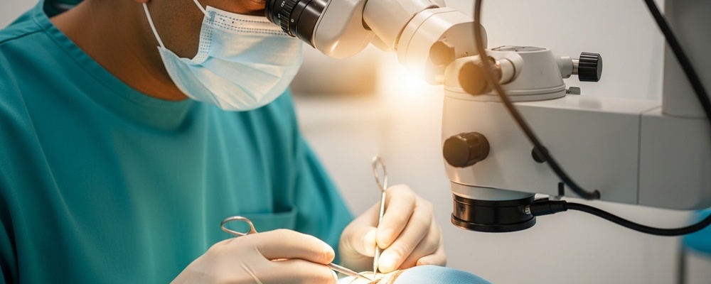
- Diseased Layer Removal: Using highly precise instruments, such as a microkeratome (a specialized blade) or an advanced laser system (like a femtosecond laser), your surgeon will carefully remove the damaged superficial layers of your cornea.
- Donor Tissue Placement: A healthy, clear donor cornea, which has been meticulously prepared and matched, is then carefully positioned onto your eye.
- Securing the Graft: The new corneal tissue is typically secured with very fine sutures, which are often thinner than a human hair. In some automated techniques, sutures might be minimized or avoided.
- Protective Measures: A protective contact lens or eye shield may be placed over your eye.
3. What to Expect Immediately After the Procedure
- Recovery Area: You will be taken to a recovery lounge where nurses will monitor you as the anesthesia wears off.
- Eye Protection: A protective eye shield or patch will be placed over your eye to prevent accidental rubbing or injury.
- Initial Vision: Your vision will likely be blurry immediately after the surgery. This is normal and will gradually improve.
- Mild Discomfort: You might experience mild discomfort, a gritty sensation, or light sensitivity. Pain medication can be prescribed if needed.
- Discharge Instructions: You will receive detailed instructions on how to care for your eye, including how to use prescribed eye drops, what activities to avoid, and when your follow-up appointment is scheduled. You will be discharged the same day.
POST-OPERATIVE CARE AND TIMELINE
Recovery and Post-Operative Care
Proper post-operative care is crucial for a successful recovery and optimal vision. Your doctor will provide specific instructions, but here’s a general guide:
Do’s:
- Use Eye Drops: Administer all prescribed eye drops (antibiotics, anti-inflammatories, lubricants) exactly as directed to prevent infection and reduce inflammation.
- Wear Eye Protection: Wear your protective eye shield or glasses, especially while sleeping or in dusty environments, for the first few weeks.
- Attend Follow-up Appointments: Regular check-ups are vital for your doctor to monitor healing, remove sutures (if necessary), and adjust your medication.
- Rest Your Eyes: Avoid prolonged reading, screen time, or activities that strain your eyes initially.
- Maintain Hygiene: Keep the area around your eye clean and avoid getting water directly into your eye during showers.
Don’ts:
- Do Not Rub Your Eye: This is extremely important to prevent dislodging the new tissue or causing injury.
- Avoid Strenuous Activities: Refrain from heavy lifting, bending, or vigorous exercise for several weeks, as these can increase eye pressure.
- No Swimming or Hot Tubs: Avoid public water sources to prevent infection.
- Do Not Wear Eye Makeup: Avoid eye makeup for at least a month to prevent irritation and infection.
- Avoid Dusty or Smoky Environments: These can irritate your healing eye.
Recovery Timeline:
- Day 1: Your vision will be blurry, and you may experience mild discomfort. Keep your eye shield on. Your first follow-up appointment is usually within 24-48 hours.
- Week 1: Vision will gradually start to improve, though it may still fluctuate. Continue using all eye drops. Avoid any activities that could put pressure on your eye.
- Month 1: Significant visual improvement is often noticeable. You’ll continue with eye drops, though the frequency might be reduced. You can gradually return to most normal activities, but avoid contact sports or anything that could impact your eye. Full vision stabilization can take several months as the eye completely heals and adjusts.
COST AND INSURANCE COVERAGE IN HYDERABAD
Cost of Superficial / Automated Lamellar Keratoplasty in Hyderabad & Insurance Options
- Estimated Local Investment: ₹60000 – ₹100000
- Cost Determinants: The final cost of Superficial / Automated Lamellar Keratoplasty in Hyderabad can vary based on several factors:
- Hospital Choice: The type of hospital (e.g., corporate chain, private clinic, government-aided) and its facilities.
- Surgeon’s Experience: Highly experienced or renowned surgeons may charge higher fees.
- Procedure Technique: Whether the procedure is performed manually or utilizes advanced automated blade-free laser processing execution platforms (like femtosecond lasers), which can increase precision and cost.
- Donor Tissue Cost: The cost associated with procuring and preparing the healthy donor corneal tissue.
- Pre-operative Diagnostics: The extent and complexity of diagnostic tests required before surgery.
- Post-operative Care: The cost of follow-up visits and prescribed medications.
- Insurance Protocol: Superficial / Automated Lamellar Keratoplasty is generally considered a therapeutic ‘Medical Necessity’ as it aims to restore or improve vision impaired by disease or injury. Therefore, it is typically covered by major TPA networks and health insurance schemes in India. However, it is crucial to explicitly outline if this is a therapeutic ‘Medical Necessity’ covered by major TPA networks and insurance schemes, or an ‘Elective/Cosmetic’ correction that typically requires out-of-pocket payment or corporate policy add-on verifications. We strongly advise contacting your insurance provider directly to understand your specific policy coverage, cashless facility availability, and any required pre-authorization procedures.
FREQUENTLY ASKED QUESTIONS
Frequently Asked Questions About Superficial / Automated Lamellar Keratoplasty
Q: Is Superficial / Automated Lamellar Keratoplasty painful?
A: The procedure is performed under local anesthesia, so you will not feel pain during the surgery. You might experience mild discomfort, a gritty sensation, or irritation in the eye during the initial recovery period, which can be managed with prescribed pain relief.
Q: How long does vision take to improve after the surgery?
A: Initial vision improvement can be noticed within several days to a few weeks. However, full visual stability and the best possible outcome typically take several months as the eye completely heals and adapts to the new corneal tissue.
Q: What is the main advantage of this procedure over a full corneal transplant?
A: The primary advantage is that it replaces only the diseased front layers of the cornea, preserving the healthy deeper layers. This often leads to faster visual recovery, a lower risk of rejection, and fewer post-operative complications compared to a full-thickness corneal transplant.
Q: Are there any risks associated with Superficial / Automated Lamellar Keratoplasty?
A: As with any surgery, there are potential risks, including infection, inflammation, graft rejection (though lower than full transplants), irregular astigmatism, and delayed healing. Your surgeon will discuss all potential risks and benefits with you.
DISCLAIMER
Disclaimer: This content is curated using artificial intelligence and may contain inaccuracies. It is provided for informational and educational purposes only and should not be considered professional medical advice. Please consult your ophthalmologist for personalized clinical care. The prices listed in this article are indicative only and may vary based on the patient’s specific condition, procedural techniques, surgical complexity, and materials used. We strongly advise discussing actual costs directly with your healthcare provider.