An ophthalmoscopy, also known as a funduscopy, is a fundamental and painless diagnostic test used to get a clear view of the back of your eye. This routine examination is crucial for assessing the health of your retina and optic nerve, allowing our specialists at Pristine Eye Hospitals to detect eye conditions in their earliest stages.
What is a Ophthalmoscopy (Funduscopy)?
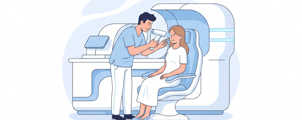
Ophthalmoscopy is a non-invasive procedure where your eye doctor uses a specialized instrument called an ophthalmoscope to look through your pupil and lens to the back of the eye. This device shines a bright light, illuminating the internal structures of your eye. This allows for a detailed examination of the fundus, which includes the retina (the light-sensitive tissue), the optic disc (where the optic nerve connects to the retina), and the network of blood vessels that nourish them.
Why is this test performed?
This essential examination is performed for several key reasons, including:
To detect and monitor diseases of the retina, such as diabetic retinopathy, macular degeneration, and retinal tears or detachments.
To evaluate the health of the optic nerve, which is critical for diagnosing and managing conditions like glaucoma or optic neuritis.
To investigate symptoms like unexplained vision loss, floaters, flashes of light, or headaches.
To identify signs of systemic health problems, such as high blood pressure or diabetes, which can manifest as changes in the eye’s blood vessels.
How to Prepare for Your Ophthalmoscopy (Funduscopy)
In most cases, minimal preparation is needed. However, to get the best possible view of your retina, your ophthalmologist may need to dilate your pupils using special eye drops.
Inform your doctor about any medications you are currently taking and if you have a history of glaucoma.
Arrange for a friend or family member to drive you home, as the dilating drops will cause blurry vision and light sensitivity for a few hours.
Bring a pair of sunglasses to wear after the appointment to help with light sensitivity.
The Procedure: What to Expect Step-by-Step
1. Your doctor or a technician may administer dilating eye drops. You will wait for about 20-30 minutes for your pupils to fully open.
2. You will be seated comfortably in a dimly lit or dark examination room to help your pupils remain dilated.
3. Your ophthalmologist will sit in front of you and use a handheld ophthalmoscope or a larger slit-lamp microscope to shine a bright light into your eye. You will be asked to look straight ahead or in different directions.
4. The doctor will carefully examine the retina, optic disc, and blood vessels in each eye. The entire examination is quick, typically lasting only 5-10 minutes.
Understanding Your Results
Immediately after the examination, your ophthalmologist will discuss the findings with you. A normal result means your retina, optic disc, and blood vessels appear healthy, with no signs of damage or disease.
If any abnormalities are detected, your doctor will explain what they mean for your vision and overall health. Findings could include signs of optic nerve damage (suggesting glaucoma), leaky blood vessels (common in diabetic retinopathy), or deposits called drusen (an early sign of macular degeneration). Based on these results, your doctor will recommend further testing or a personalized treatment plan to protect your sight.
Frequently Asked Questions
Is an ophthalmoscopy painful?
No, the test is completely painless. You may experience mild, temporary discomfort from the bright light used during the exam.
How long does the test take?
The examination itself is very quick, usually taking less than 10 minutes for both eyes. However, if your pupils are dilated, you should plan to be at the hospital for about an hour to allow time for the drops to work.
Will my vision be blurry after an ophthalmoscopy?
If dilating drops are used, your vision will be blurry and you will be sensitive to light for several hours afterward. For this reason, it is not safe to drive until the effects have worn off completely.