Gonioscopy is a fundamental, painless eye examination that allows your ophthalmologist to get a clear view of your eye’s internal drainage system. This quick diagnostic test is crucial for accurately diagnosing and managing glaucoma. At Pristine Eye Hospitals, we use this procedure to determine the specific type of glaucoma you may have, ensuring you receive the most effective treatment.
What is a Gonioscopy?
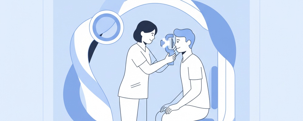
A Gonioscopy is an in-office procedure that uses a special contact lens with mirrors, called a goniolens or gonioscope, to inspect the angle where the iris meets the cornea. This “drainage angle” contains the trabecular meshwork, a tiny structure responsible for draining fluid (aqueous humor) out of the eye. By examining this angle, your doctor can determine if it is open or closed, which is the key difference between the two main types of glaucoma.
Why is this test performed?
Your ophthalmologist may recommend a gonioscopy for several important reasons, including:
To differentiate between open-angle glaucoma and angle-closure glaucoma, as their treatments are very different.
To check for any blockages, scarring, or damage to the eye’s drainage system resulting from trauma or previous eye surgery.
To identify abnormal blood vessels or tumors that could be affecting eye pressure.
To monitor the effectiveness of certain glaucoma treatments or surgical procedures.
How to Prepare for Your Gonioscopy
This is a straightforward diagnostic test that requires very little preparation. However, to ensure the process is smooth and comfortable, please keep the following in mind:
Inform your doctor about all medications you are currently taking and any known allergies.
If you wear contact lenses, you will need to remove them before the test begins.
It is often helpful to have someone accompany you, although driving is usually permissible as your pupils are not typically dilated for this specific test.
The Procedure: What to Expect Step-by-Step
The entire gonioscopy procedure is quick, typically lasting only a few minutes. Here is what you can expect:
1. Numbing the Eye: Your doctor will first apply anaesthetic eye drops to completely numb the surface of your eye, ensuring you feel no pain.
2. Positioning: You will be seated comfortably and asked to place your chin and forehead on the rests of a slit lamp (a specialised microscope for eye exams).
3. Placing the Lens: Your doctor will gently place the small, smooth goniolens onto the front surface of your eye. You may feel a slight pressure, but no pain.
4. Examination: Using the slit lamp’s light and magnification, the doctor will carefully rotate the lens to view the entire 360-degree drainage angle and assess its structure.
Understanding Your Results
Your ophthalmologist at Pristine Eye Hospitals will discuss your results with you immediately after the test. The primary finding is the state of your drainage angle. An “open angle” means the drainage pathway is physically unobstructed, which is characteristic of open-angle glaucoma. In this case, the drainage system itself is not working efficiently.
If the angle is determined to be “narrow” or “closed,” it indicates that the iris is blocking fluid from reaching the drainage system. This finding is characteristic of angle-closure glaucoma, which can sometimes be a medical emergency. Understanding your angle’s structure is the first step in creating a personalised treatment plan to preserve your vision.
Frequently Asked Questions
Is a gonioscopy painful?
No, the test is not painful. The anaesthetic eye drops completely numb the surface of your eye, so you will only feel a light touch or pressure from the lens.
How long does a gonioscopy take?
The examination is very quick and efficient. It typically takes less than five minutes to examine both eyes thoroughly.
Are there any side effects?
Side effects are rare and minimal. Some patients may experience temporary blurry vision or slight irritation for a short while after the numbing drops wear off, but this resolves quickly.