While a standard eye chart measures your ability to see black letters on a white background, real-world vision is much more complex. Contrast sensitivity testing evaluates your ability to distinguish between objects and their background in various lighting conditions. This provides a more complete and accurate picture of your true functional vision.
What is a Contrast Sensitivity Testing?
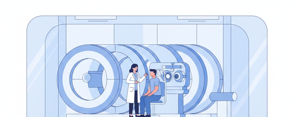
Contrast sensitivity testing is a non-invasive diagnostic exam that measures how well your eyes can detect subtle differences in shading and patterns. Unlike a standard visual acuity test that uses high-contrast black letters, this test utilizes specialized charts, such as the Pelli-Robson chart, which display letters or patterns in progressively lighter shades of grey. This allows our ophthalmologists to quantify your ability to see in low-contrast environments, which is crucial for activities like driving at night, navigating in fog, or seeing facial expressions clearly.
Why is this test performed?
This test is essential for a comprehensive understanding of your visual function and is performed to:
Detect early signs of eye conditions like cataracts, glaucoma, macular degeneration, and diabetic retinopathy, which can reduce contrast sensitivity before affecting visual acuity.
Monitor the progression of known eye diseases and the effectiveness of their treatment.
Evaluate the quality of your vision after refractive surgery (like LASIK) or cataract surgery with premium intraocular lens implants.
Assess vision for specific tasks requiring good low-contrast perception, such as driving at night or recognizing objects in dim light.
How to Prepare for Your Contrast Sensitivity Testing
Preparing for a contrast sensitivity test is simple and requires no special effort. To ensure the most accurate results, we recommend following these straightforward guidelines:
Bring your current prescription glasses or contact lenses with you to the appointment.
Inform your ophthalmologist about any existing eye conditions, recent eye surgeries, or medications you are taking.
Ensure you are well-rested, as fatigue can sometimes affect visual performance.
The Procedure: What to Expect Step-by-Step
1. You will be comfortably seated at a specific, standardized distance from a specialized chart or a computer screen.
2. The chart will display a series of letters or patterns (such as wavy lines or circles) in various shades of grey against a uniform background.
3. You will be asked to identify the letters or describe the patterns, which will become progressively fainter and harder to distinguish. Each eye may be tested separately.
4. Your responses are carefully recorded to determine the faintest contrast level you can reliably see, providing a precise measurement of your contrast sensitivity.
Understanding Your Results
Your results will be plotted on a graph, creating a “contrast sensitivity curve” that shows how well you can perceive contrast across different levels of detail. A healthy visual system will fall within a normal range, while a score below this range indicates reduced contrast sensitivity. This can be an early indicator of an underlying eye health issue, even if your vision on a standard eye chart is 20/20.
Our expert ophthalmologists at Pristine Eye Hospitals will carefully review the results with you, explaining what they mean for your overall eye health. Based on these findings, they may recommend further diagnostic tests or discuss potential treatment options to address the underlying cause and improve your quality of vision.
Frequently Asked Questions
Is the contrast sensitivity test painful?
No, the test is completely non-invasive and painless. It feels very similar to reading a standard eye chart, just with images and letters of varying faintness.
How is this different from a regular eye chart test?
A regular chart measures visual acuity (sharpness) with high-contrast symbols. Contrast sensitivity testing provides a more detailed assessment of your functional vision by measuring your ability to see in realistic, low-contrast situations.
How long does the test take?
The test itself is very quick and efficient. It typically takes only 5 to 10 minutes to complete and is often performed as part of a comprehensive eye examination.