The Anterior Segment OCT (Angle) is a quick, painless, and non-invasive imaging test that provides a detailed, cross-sectional view of the front part of your eye. At Pristine Eye Hospitals, we use this advanced technology to precisely measure the eye’s drainage angle, a critical factor in assessing the risk of certain types of glaucoma. This powerful diagnostic tool helps our specialists protect your vision with early and accurate detection.
What is an Anterior Segment OCT (Angle)?
Optical Coherence Tomography (OCT) is an imaging technology that uses light waves to capture high-resolution, microscopic images of your eye’s structures. The Anterior Segment OCT specifically focuses on the front section of the eye, including the cornea, iris, and lens. This particular test is used to visualize and measure the iridocorneal angle—the “drainage angle” where the iris meets the cornea—which is responsible for draining fluid from the eye.
Why is this test performed?
Your ophthalmologist at Pristine Eye Hospitals may recommend this test for several key reasons:
To accurately assess your risk for angle-closure glaucoma, a condition where the drainage angle becomes blocked, causing a sudden increase in eye pressure.
To visually inspect and measure the anatomy of the drainage angle in a non-contact way, which is more detailed than traditional methods.
To monitor changes in the angle over time, especially for patients being managed for narrow angles or glaucoma.
To plan for and evaluate the outcomes of procedures like laser iridotomy or cataract surgery, which can affect the angle’s structure.
How to Prepare for Your Anterior Segment OCT (Angle)
This is a straightforward and simple procedure that requires minimal preparation. Generally, no special steps like fasting or pupil dilation are needed, allowing us to view the angle in its natural state.
Please inform your technician about any eye drops you are currently using or any known eye conditions.
If you wear contact lenses, you may be asked to remove them just before the test begins.
Relax, as the test is quick, non-contact, and completely painless.
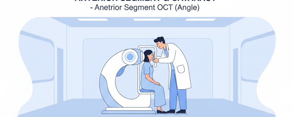
The Procedure: What to Expect Step-by-Step
1. You will be seated comfortably in front of the OCT machine.
2. You will be asked to place your chin on a chin rest and your forehead against a support bar. This helps keep your head perfectly still for clear images.
3. The technician will instruct you to look at a small target light inside the machine.
4. The scanner will quickly capture detailed images of your eye’s angle without ever touching it. The entire scan for both eyes is typically completed in just a few minutes.
Understanding Your Results
The images from the Anterior Segment OCT provide your ophthalmologist with precise, objective measurements and a clear visualization of your eye’s drainage angle. Your doctor will analyze these images to determine if your angle is open, narrow, or closed. A narrow angle suggests a higher risk for angle-closure glaucoma, as it can impede the natural outflow of fluid from the eye.
Based on these findings, your doctor can determine if you require monitoring, preventive treatment like a laser iridotomy, or other management strategies. This early and accurate diagnosis is crucial for preserving your long-term vision and preventing optic nerve damage associated with glaucoma.
Frequently Asked Questions
Is the Anterior Segment OCT test painful?
No, the test is completely non-contact and painless. The scanner uses light to capture images and nothing will touch your eye during the procedure.
How long does the test take?
The test is very fast. While you may be in the room for a few minutes for positioning, the actual image capture for each eye takes only a few seconds.
Will my pupils be dilated for this test?
Typically, pupil dilation is not required for an Anterior Segment OCT (Angle) test. This allows the ophthalmologist to evaluate the drainage angle in its natural, undilated state.