
Keratoconus and Corneal Ectasia
Mar 15, 2024

What is Keratoconus?
Keratoconus is a progressive eye condition where the cornea—the transparent, dome-shaped outer layer of the eye—gradually thins and bulges outward into a cone-like shape.
This results in blurry or distorted vision, increased sensitivity to light, and, if left untreated, can lead to vision loss.The onset of keratoconus typically occurs during puberty or late teens and early 30s, with gradual progression over a decade or more. Both eyes are usually affected, though one eye may experience more severe symptoms than the other.
Patients with keratoconus often experience stress and anxiety due to the impact on their quality of life. While keratoconus is a chronic condition that cannot be fully cured, the goodnews is that most cases can be effectively managed. With early diagnosis, advanced treatment options, and expert care, patients with keratoconus can maintain an active, healthy lifestyle.
Video: What is Keratoconus? Explained by renowned surgeon and founder of Pristine Eye Hospitals, Dr C Jagadesh Reddy
Keratoconus Symptoms & Signs
Symptoms and their intensity vary depending on the stage of the condition—whether in early or advanced stages.
Blurred vision
Frequent changes in eye power
Irritation or itching in the eye
Sudden worsening of vision
Distorted or double vision
Astigmatism
Sensitivity to light
Difficulty seeing in dim light or at night
Halos or glares around light
Fleischer ring (brown or greyish-green rings around the cornea)
Keratoconus Causes
While the exact cause of keratoconus remains unknown, several risk factors are associated with its development:
Frequent or excessive eye rubbing
Use of poorly fitting contact lenses or improper lens care
Family history of keratoconus (about 1 in 10 people with keratoconus have a parent with the condition)
Chronic eye inflammation/Eye Allergy
Corneal scarring or damage due to eye injury
Severe eye complications
Congenital disorders (eye conditions present at birth)
Genetic factors, including Down Syndrome
Loss of collagen in the cornea (collagen is the protein that makes up the middle layer of the cornea)
Keratoconus Diagnosis
Keratoconus is diagnosed through a comprehensive eye examination, corneal assessment, and a review of family and medical history. Some of the diagnostic tests include:
Slit-Lamp Examination and Refraction Test: A slit-lamp examination closely examines the cornea, and an eye refraction test evaluates the eye’s refractive (spectacle) power.
Corneal Topography/Tomography: This non-invasive imaging technique maps the curvature of the cornea and is one of the most accurate tests for diagnosing keratoconus and monitoring progression.
Pachymetry: This test measures the thickness of the cornea, which is essential for assessing keratoconus progression.
Optical Coherence Tomography (OCT): This is another non-invasive imaging technique that produces high-quality images to assist in the detection and monitoring of the progression of keratoconus.
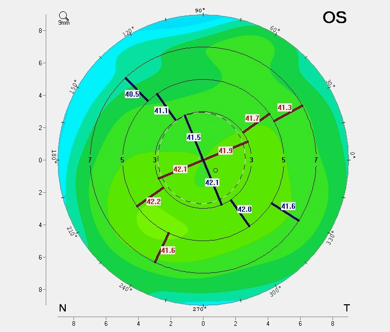
Image: Pentacam AXL wave Scan of a Healthy Cornea – Normal Eye Shape Comparison
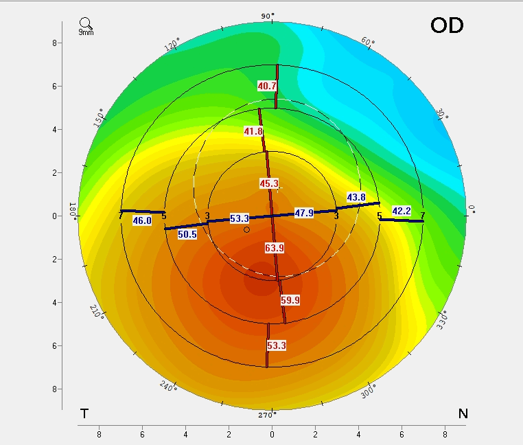
Image: Pentacam AXLwave Scan Showing Keratoconus – Irregular Corneal Shape Detection
Keratoconus Treatment and Management
While keratoconus cannot be fully cured, it can be effectively managed through various treatment options depending on the severity of the condition. Your ophthalmologist will recommend the most suitable treatment based on the severity of the condition and lifestyle.
Glasses or Contact Lenses: In the early stages, glasses or soft contact lenses are prescribed for vision correction.
Specialty Contact Lenses: As keratoconus progresses, specialty contact lenses such as scleral lenses, PROSE lenses, or rigid gas permeable (RGP) lenses may be recommended.
Corneal Cross-Linking (CXL): This minimally invasive procedure is performed to slow the progression of keratoconus by stabilising the cornea, preventing further thinning and bulging. The process involves applying riboflavin (vitamin B2) drops to the cornea, followed by exposure to ultraviolet light using a specialised machine. While CXL does not directly improve vision, it reduces the need for a corneal transplant.
Advanced Sub-400 Micron Protocols for Cross-Linking: Pristine Eye Hospitals specializes in advanced sub-400 micron protocols for collagen cross-linking, making it possible to treat very thin corneas effectively.
Intracorneal Ring Segments (ICRS): These small, crescent-shaped implants are inserted into the cornea to flatten it and reduce the cone-shaped bulge, thereby improving vision.
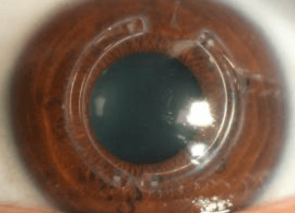
Image: Keratoconus Treatment with INTACS Ring – Corneal Support for Better Vision
Topography-guided Photorefractive Keratectomy (Topography-guided PRK or TG-PRK): This laser procedure uses Contoura laser technology to reshape the cornea and improve vision in patients with keratoconus.
Corneal Allogenic Intrastromal Ring Segments (CAIRS): This innovative procedure involves implanting allogenic tissue within the cornea to improve corneal strength and stability.
Corneal Transplant (Keratoplasty): In advanced stages, where the cornea becomes extremely thin or vision can no longer be corrected through other means, a corneal transplant may be required. The damaged cornea is replaced with a healthy donor cornea. Depending on the case, either a full-thickness transplant (penetrating keratoplasty) or a partial-thickness transplant (deep anterior lamellar keratoplasty, or DALK) may be recommended

Image: Clear Corneal Transplant with Sutures – Post-Keratoplasty Recovery
Research and Innovation in Keratoconus Detection
Dr. C. Jagadesh Reddy, a leading cornea surgeon at Pristine Eye Hospitals, has been practicing keratoconus management for over 20 years. His research focuses on AI-based techniques for the early detection of keratoconus progression.
For further reading on Dr. Reddy’s research:
1. Reddy JC, et al. KEDOP: Keratoconus early detection of progression using tomography images. Eur J Ophthalmol. 2022;32:2554-2564. https://pubmed.ncbi.nlm.nih.gov/35343267/
2. Das AV, Deshmukh RS, Reddy JC, et al. Keratoconus in India: Clinical presentation and demographic distribution based on big data analytics. Indian J Ophthalmol. 2024 Jan 1;72:105-110. https://pubmed.ncbi.nlm.nih.gov/38131579/
3. Tharini B, Sahebjada S, Borrone MA, Vaddavalli P, Ali H, Reddy JC. Keratoconus in pre-teen children: Demographics and clinical profile. Indian J Ophthalmol. 2022;70:3508-3513. https://pubmed.ncbi.nlm.nih.gov/36190036/
Keratoconus Care at Pristine Eye Hospitals
Pristine Eye Hospitals has a specialised team with expertise in managing keratoconus patients and offers the latest diagnostic tests for early detection. Our compassionate team of ophthalmologists, optometrists, and technicians provides personalized treatment plans, including advanced surgical care options, to help stabilise the condition and improve patients' quality of life.